Exploring the Benefits of 3D Conebeam Imaging
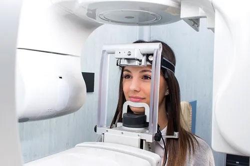
Advancements in dental imaging technology have significantly enhanced the diagnostic capabilities available to dental professionals. One such innovation is 3D Conebeam Imaging, a cutting-edge technique that provides comprehensive and detailed images of dental structures. This method is increasingly becoming a staple in modern dental practices due to its accuracy, efficiency, and ability to improve patient outcomes.
What is 3D Conebeam Imaging?
3D Conebeam Imaging, also known as Cone Beam Computed Tomography (CBCT), is a specialized type of x-ray equipment used when traditional dental x-rays are not sufficient. This technology provides a 3D view of the teeth, soft tissues, nerve pathways, and bone in a single scan, offering a more complete picture than conventional imaging methods.
Benefits of 3D Conebeam Imaging
Enhanced Diagnostic Accuracy
3D Conebeam Imaging offers unparalleled diagnostic accuracy. This technology allows for a more comprehensive evaluation of complex dental conditions. The benefits include:
- Detailed visualization of hard and soft tissues.
- Precise assessment of bone structure and quality.
- Improved detection of hidden dental issues such as root fractures or impacted teeth.
Improved Treatment Planning
The detailed imagery provided by 3D Conebeam Imaging facilitates precise treatment planning. For dental procedures, understanding the exact positioning and condition of teeth and surrounding structures is crucial. This technology enhances planning by:
- Providing detailed images that aid in the accurate placement of implants.
- Helping in the assessment of airway passages for orthodontic procedures.
- Assisting in the evaluation of temporomandibular joint disorders.
For more on how this technology is applied in various treatments, visit this 3d pano conebeam resource.
Reduced Radiation Exposure
Compared to traditional CT scans, 3D Conebeam Imaging significantly reduces the patient’s exposure to radiation. This is particularly beneficial for children or patients requiring multiple scans over time. Key points include:
- Lower radiation doses compared to conventional CT scans.
- Safe and effective for regular monitoring and follow-up.
- Suitable for detailed assessments without compromising patient safety.
Applications in Modern Dentistry
The versatility of 3D Conebeam Imaging makes it applicable in various fields of dentistry. Some common applications include:
Implant Dentistry
One of the primary uses of 3D Conebeam Imaging is in implant dentistry, where precision is crucial. The technology aids in:
- Determining the optimal placement for implants.
- Evaluating the density and volume of the jawbone.
- Reducing the risk of complications during implant surgery.
Orthodontics
In orthodontics, 3D imagery helps in the comprehensive evaluation of the patient’s dental and skeletal structure. This assists in:
- Designing precise orthodontic treatment plans.
- Monitoring the progress of tooth movement.
- Predicting jaw growth patterns in young patients.
Endodontics
This imaging technology is invaluable in endodontics for diagnosing complex cases, including:
- Identifying root fractures or canal obstructions.
- Planning endodontic surgeries with minimal invasiveness.
- Evaluating the success of root canal treatments.
Conclusion
3D Conebeam Imaging represents a significant advancement in dental imaging technology, offering numerous benefits over traditional methods. Its ability to provide detailed, accurate, and comprehensive views of dental anatomy makes it an indispensable tool in modern dentistry. As dental care continues to evolve, the adoption of such advanced technologies will play a crucial role in improving patient outcomes and enhancing the precision of dental treatments. For more insights into the possibilities offered by 3D Conebeam Imaging, explore the latest resources and advances in dental imaging.